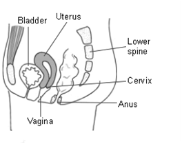
Transvaginal Ultrasonography
- The ultrasonography is performed transvaginally on empty bladder as it gives much clearer images.
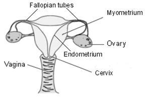
- The scan will evaluate the size, shape and condition of the internal reproductive organs like womb (uterus), inner lining of the womb (endometrium), both side eggs pouch (ovaries) and the adjacent pelvic area (adnexa).
- Transvaginal ultrasonography is a very important diagnostic modality and can evaluate the condition of the internal reproductive organs as follows –
- Examining the size and shape of the uterus,
- Presence of fibroid or polyp or cyst or septum or adhesions or tumor in the uterus,
- Presence mullerian anomalies (genetically abnormal shape of the uterus),
- Examining the number and size of follicles and the general egg reserve of the ovaries,
- Presence of cyst in the ovaries (polycystic ovaries),
- Detecting ectopic pregnancy (pregnancy in the fallopian tube or in the ovary)
- Examining the thickness and stage of development of the endometrium,
- Abnormal unhealthy or polypoidal endometrium.
Follicular Study
- Follicular study (ovulation tracking) involves tracking of the follicles (fluid containing sac with eggs) of both the ovaries in a reproductive cycle. The follicle number and measure in mm of both the ovaries are observed carefully.
- Follicular study is an important modality to evaluate how the egg is developing and/or responding to the treatment. Hormonal injection may be required for the development of egg.
- It is done frequently like on day 2- day3, day 7 to day 9, day 11 of the menstrual cycle or as per required.